Neodent · Short Zygomatic

Neodent / Zygoma S

Short-body zygomatic implant for cases with less severe maxillary atrophy. Engages the zygomatic process with a reduced length profile, enabling the extrasinus and ZAGA technique with lower surgical complexity.

Short Zygomatic 30–40 mm · 5 Lengths Moderate Atrophy Grand Morse® Extrasinus Technique Fixed Rehab
Pricing
Available on request · Contact for current pricing and availability
Product
Zygoma S · 5 Lengths · 30–40mm

Product Overview

Implant ClassificationShort Zygomatic Implant — zygomatic cortical anchorage at 30–40 mm depth
Primary IndicationModerate posterior maxillary atrophy — zygomatic process accessible without full-length trajectory
Apex DesignConical apex with spherical self-tapping tip and helical flutes for cortical purchase
Thread DesignProgressive trapezoidal threads — denser pitch at apical zone for dense zygomatic bone
Surface TreatmentNeoPoros — sandblasted + acid-etched (SLA) on full length except smooth cervical zone
DiameterØ 4.0 mm
Available Lengths30 / 32.5 / 35 / 37.5 / 40 mm (5 options)
ConnectionGrand Morse® 16° internal cone — angled cervical emergence
Preferred TechniqueExtrasinus / ZAGA 0–2 — lateral wall path, no sinus penetration required
Immediate LoadingSupported when primary stability criteria are met (torque ≥35 N.cm, ISQ ≥65)
Prosthetic CompatibilityFull Neodent GM system: MUA 0°/17°/30°, Ti-Base, digital workflows
MaterialGrade IV/V titanium alloy

What is the Zygoma S?

A purpose-built bridge between conventional implants and full-length zygomatic systems — calibrated for the moderately atrophic maxilla.

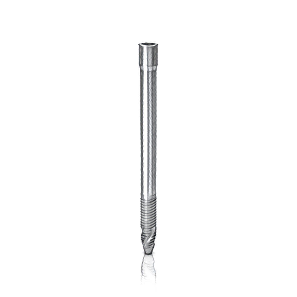
The Neodent Zygoma S was developed to address a gap that had long existed in zygomatic implantology: patients with moderate posterior maxillary atrophy — significant enough to preclude conventional implants, yet not so severe as to require the 45–55 mm trajectory of a standard zygomatic implant. In these patients the zygomatic process is accessible at shorter depths (30–40 mm), allowing anchorage with a reduced-length body. The Zygoma S exploits precisely this anatomy, delivering the biomechanical security of zygomatic cortical engagement with less surgical complexity, a smaller osteotomy, and a reduced risk profile compared to the full Zygoma GM.

The defining clinical advantage of the Zygoma S is its alignment with the extrasinus technique and the ZAGA (Zygomatic Anatomy Guided Approach) classification system. In the extrasinus approach — ZAGA classes 0 through 2 — the implant body travels along the outer (lateral) surface of the posterior maxillary wall, completely bypassing the maxillary sinus. This eliminates sinus-related complications such as chronic sinusitis, oroantral communication, and the management burden of a trans-sinus implant body. The shorter length of the Zygoma S is geometrically aligned with the bone available in this extrasinus path: a shallower, more lateral trajectory that still achieves full engagement of the zygomatic process cortex. It is this combination — shorter trajectory, no sinus penetration, and dense cortical anchorage — that defines the Zygoma S clinical niche.

From a prosthetic standpoint, the Zygoma S connects via the Grand Morse® 16° cone, identical to the full-length Zygoma GM and the entire Neodent GM implant family. This compatibility means Multi-Unit Abutments (MUAs), Ti-Bases, digital scan bodies, and CAD/CAM workflows already used in the practice transfer directly to Zygoma S cases. The angled cervical emergence accommodates zygomatic anatomy, while the appropriate MUA angle (0°, 17°, or 30°) corrects the prosthetic platform to a parallelism usable for full-arch screw-retained prostheses without any additional custom components.

Clinically, the Zygoma S is most indicated in bilateral cases of moderate atrophy, where one Zygoma S per posterior quadrant is combined with two anterior conventional Grand Morse implants in an All-on-4 or All-on-6 configuration. It is also the implant of choice when a patient has refused sinus augmentation, when prior sinus lifts have failed, or when conventional posterior implant placement is anatomically impossible. Because zygomatic cortical bone is dense and naturally offers high primary stability regardless of maxillary ridge quality, the Zygoma S frequently supports same-day loading protocols — one of its most clinically valued features.

Zygoma S vs. Zygoma GM: Key Differences

Zygoma S (This Product)
Lengths 30–40 mm, 5 options
Moderate maxillary atrophy
Extrasinus / ZAGA 0–2
No sinus penetration needed
Shorter, simpler surgery
Ideal for graft refusal cases
Zygoma GM (Full Length)
Lengths 30–55 mm, 10 options
Severe / complete maxillary loss
Intrasinus + extrasinus ZAGA 0–4
Trans-sinus classic technique
Greater apical depth required
Quad zygoma for fully edentulous

Engineering Behind the Zygoma S

Every design element is optimized for the extrasinus trajectory and the cortical bone of the zygomatic process.

📐
Compact Length Profile — 30 to 40 mm
Five precisely calibrated lengths in 2.5 mm increments allow the surgeon to match the exact zygomatic depth without over-penetrating the arch. CBCT planning determines the optimal length before surgery, eliminating guesswork at the operating table.
🏗
Extrasinus / ZAGA Architecture
The implant body geometry and entry angle are calibrated for a lateral-wall extrasinus path. The Zygoma S courses along the outer cortex of the maxilla, completely bypassing the sinus membrane and eliminating the primary source of zygomatic implant complications.
🔗
Grand Morse® 16° Cone Connection
Deep internal cone engagement eliminates micro-movement, bacterial infiltration, and the microgap at the implant-abutment interface. The same connection as all Neodent GM implants — full prosthetic ecosystem compatibility from day one of adoption.
🧬
NeoPoros SLA Surface
Sandblasted and acid-etched surface with proven osteoconductive micro-topography. Accelerates osseointegration in the dense zygomatic cortex, supporting aggressive loading timelines. Surface extends the full length of the implant body except the smooth cervical collar.
🛡
Tissue Protect Cervical Zone
Smooth machined collar at the trans-mucosal emergence zone reduces crestal stress concentration and supports healthy soft tissue adaptation. Particularly important in zygomatic cases where the peri-implant mucosa spans a longer distance from bone to emergence.
Immediate Loading Protocol
Zygomatic bone is naturally dense and delivers high primary stability. When insertion torque and ISQ values meet protocol thresholds (≥35 N.cm, ISQ ≥65), the Zygoma S supports same-day provisional loading — enabling fixed teeth on the day of surgery in qualifying patients.

When to Choose the Zygoma S

The Zygoma S occupies a distinct niche between standard implants and the full Zygoma GM — precise patient selection is the key to success.

Moderate Posterior Maxillary Atrophy

Patients with significant posterior ridge resorption but sufficient anatomy for a 30–40 mm zygomatic trajectory. CBCT confirms zygomatic process accessibility at this depth with a compatible lateral wall profile (ZAGA 0–2).

ZAGA 0, 1, and 2 Classification

The ZAGA classification system identifies patients whose lateral wall morphology supports an extrasinus implant path. Classes 0–2 — concave-to-flat lateral wall — are the ideal indication for the Zygoma S. Class 3 and 4 require a full-length approach.

Patients Declining Bone Grafts

For patients who have refused sinus lifts or alveolar augmentation — or where prior grafting has failed — the Zygoma S provides a graft-free posterior anchor in native zygomatic bone, bypassing the compromised maxillary ridge entirely.

All-on-4 / All-on-6 Posterior Anchors

When posterior conventional implant placement is impossible due to bone volume or sinus proximity, Zygoma S implants serve as the posterior pillars of a full-arch fixed rehabilitation combined with anterior conventional Grand Morse implants.

Bilateral Symmetric Moderate Atrophy

One Zygoma S per posterior quadrant provides symmetric bilateral posterior support for a full-arch prosthesis — a balanced configuration that distributes occlusal forces predictably and simplifies prosthetic planning.

Failed Conventional Posterior Implants

When posterior implants have failed due to compromised bone quality or proximity to the sinus floor, the Zygoma S offers a rescue solution anchored in the reliable cortical bone of the zygomatic process, avoiding the problem site entirely.

Extrasinus Technique: Clinical Workflow

Key steps and considerations for planning and placing the Zygoma S using the extrasinus ZAGA approach.

1

CBCT Planning and ZAGA Classification

Pre-operative CBCT is mandatory. Import DICOM data into planning software (coDiagnostiX, Simplant, or equivalent) to measure zygomatic process depth, confirm lateral wall morphology, and classify the patient as ZAGA 0–2 for extrasinus compatibility. Confirm the Zygoma S length range (30–40 mm) matches the planned trajectory. If the anatomy requires deeper penetration, the Zygoma GM is indicated instead.

2

Surgical Access and Lateral Wall Exposure

Elevate a full-thickness mucoperiosteal flap in the posterior maxilla to expose the lateral wall of the maxilla and the base of the zygomatic process. Clear visualization of the lateral cortex is essential — the extrasinus technique routes the implant along this surface without entering the sinus space. Use a surgical guide when available to verify entry angle and depth.

3

Osteotomy Along Lateral Cortex

Using the dedicated Zygoma drill sequence, create a channel along the lateral cortex of the maxilla at the planned trajectory. The drill follows the outer bone surface without creating a sinus window — a defining feature of the extrasinus technique. Drill to the exact length of the selected implant (30, 32.5, 35, 37.5, or 40 mm) to maximize cortical thread engagement in the zygomatic process.

4

Implant Insertion and Primary Stability Verification

Insert the Zygoma S using the integrated holder at the planned angle. Advance until the smooth collar reaches the planned mucosal level. Measure insertion torque with a calibrated wrench. Primary stability in zygomatic cortical bone typically exceeds 35 N.cm. Record ISQ via resonance frequency analysis if available. Values ≥65 ISQ support immediate loading eligibility.

5

MUA Placement and Prosthetic Platform Creation

Seat the appropriate Grand Morse Multi-Unit Abutment at 32 N.cm — selecting 0°, 17°, or 30° based on implant angulation relative to the planned prosthetic axis. MUAs create the parallelism necessary for full-arch prosthetic delivery. Take a digital intraoral scan or conventional open-tray impression of all MUA positions.

6

Provisional Delivery and Follow-Up Protocol

If loading criteria are met, deliver the screw-retained provisional prosthesis on the day of surgery. The provisional should be in light occlusal contact — no lateral excursive load during the osseointegration phase. Monitor at 2 weeks, 6 weeks, and 3 months. Definitive full-arch prosthesis (acrylic, hybrid, or zirconia) is delivered after confirmed osseointegration, typically at 3–6 months post-operatively.

Available Lengths and Configurations

All Zygoma S sizes share Ø 4.0 mm diameter and Grand Morse® 16° connection. Length selection is driven by CBCT-measured zygomatic trajectory depth.

30mm
32.5mm
35mm
37.5mm
40mm
Length Diameter Connection ZAGA Compatibility Clinical Note
30 mmØ 4.0 mmGrand Morse® 16°ZAGA 0–1Most conservative option — minimal zygomatic penetration for near-adequate anatomy
32.5 mmØ 4.0 mmGrand Morse® 16°ZAGA 0–1Additional 2.5 mm of zygomatic cortical purchase for improved primary stability
35 mmØ 4.0 mmGrand Morse® 16°ZAGA 1–2Standard Zygoma S length — most frequently selected for moderate atrophy
37.5 mmØ 4.0 mmGrand Morse® 16°ZAGA 1–2Greater zygomatic engagement — for more advanced posterior resorption within Zygoma S range
40 mmØ 4.0 mmGrand Morse® 16°ZAGA 2Maximum Zygoma S length — at the transition point to Zygoma GM territory for ZAGA 2 anatomy

Frequently Asked Questions

How is the Zygoma S different from the Zygoma GM?
The Zygoma S offers lengths of 30–40 mm (5 options) and is specifically designed for extrasinus placement via the ZAGA 0–2 lateral wall approach, with no sinus penetration required. The Zygoma GM extends to 55 mm (10 options) and accommodates all ZAGA classes including intrasinus trajectories for severe atrophy. When zygomatic cortical bone is accessible at under 40 mm depth and the lateral wall morphology is extrasinus-compatible, the Zygoma S is preferred for its reduced surgical complexity and lower sinus-related risk profile.
Does the extrasinus technique work for all patients?
No — patient selection requires CBCT evaluation and formal ZAGA classification. Patients classified as ZAGA 3 or 4 (very convex or protruding lateral wall configurations) are not suitable for the extrasinus approach and require either an intrasinus technique with a longer implant or a combined protocol. ZAGA 0–2, which represents the majority of zygomatic candidates with moderate atrophy, is extrasinus-compatible and appropriate for the Zygoma S.
Can the Zygoma S be immediately loaded?
Yes — immediate loading is possible when adequate primary stability is achieved. The dense cortical bone of the zygomatic process typically provides insertion torque ≥35 N.cm and ISQ values ≥65, meeting most immediate loading protocols. When these thresholds are not met, a delayed loading protocol of 8–12 weeks is indicated. The surgeon must assess each case individually and never place a provisional prosthesis on an implant that did not achieve adequate primary stability.
What prosthetic components are compatible with the Zygoma S?
The Zygoma S uses the Grand Morse® 16° connection — identical to the entire Neodent GM implant line. It is fully compatible with Neodent GM Multi-Unit Abutments (0°, 17°, 30°), GM Ti-Bases, GM prosthetic bars, and all standard Grand Morse digital workflow components. Clinicians and laboratories already working with Neodent GM have immediate access to the complete prosthetic ecosystem for Zygoma S cases without adding any new components.
Is special training required to place the Zygoma S?
Yes — zygomatic implant surgery requires advanced training beyond standard implantology, even with the shorter Zygoma S. The proximity to the orbit, maxillary sinus, and infraorbital nerve demands thorough CBCT-guided pre-operative planning and anatomical expertise. Surgeons should complete dedicated zygomatic implant training courses and, ideally, perform the procedure under mentorship before independent placement. Neodent offers hands-on cadaver training and a structured Zygoma certification curriculum.

Complete the Full-Arch Protocol

The Zygoma S integrates seamlessly with the Grand Morse ecosystem. These components complete your surgical and prosthetic workflow.

Request Zygoma S Pricing

Contact our team for current stock, pricing, and clinical support. We supply Neodent GM components with fast shipping and pre-sales consultation.